Hip Preservation Case Examples
Pre-Operative & Post-Operative Photos
Actual Cases performed by Our Surgeons
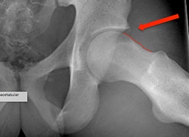
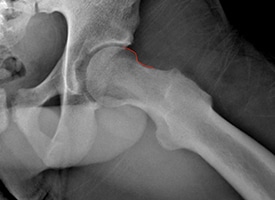
Re-Contouring
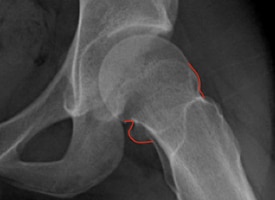
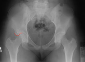
The red line demonstrates a large residual CAM deformity after in situ screw fixation of Slipped Capital Femoral Epiphysis (SCFE). This deformity limits range of motion and impinges against the hip socket causing damage to the joint.
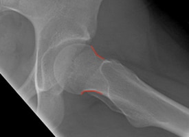
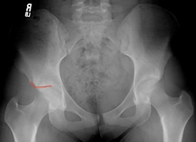
The red line demonstrates re-contouring of the femoral neck after arthroscopic treatment. This treatment increases range of motion and prevents further damage to the hip.
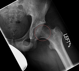
This x-ray demonstrates a large CAM deformity on the femoral neck that can impinge and damage the hip socket.
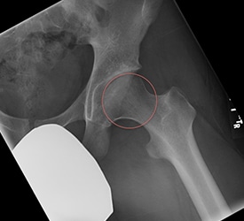
This x-ray demonstrates re-contouring of the femoral neck after arthroscopic treatment to eliminate impingement and preserve the hip joint.
This x-ray demonstrates Legg Calve Perthes disease with an abnormally shaped femoral head causing impingement. This can limit range of motion and damage the hip joint.
This x-ray demonstrates arthroscopic re-contouring of the Legg Calve Perthes deformity. The ball of the hip now lies within the circle. This treatment can increase range of motion and preserve the hip joint.
Periacetabular Osteotomy
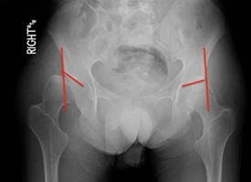
This x-ray demonstrates right hip dysplasia and a right hip dislocation. Notice the upward sloping hip socket and position of the right hip outside of edge of the socket as demonstrated by the vertical red line.
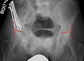
This x-ray demonstrates improved position of the hip after periacetabular osteotomy. Notice that the hip slope is improved and the ball of the hip joint is now inside the vertical line.
Realignment
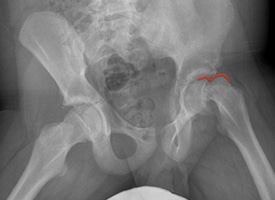
This x-ray demonstrates a severe unstable Slipped Capital Femoral Epiphysis (SCFE) of the left hip.
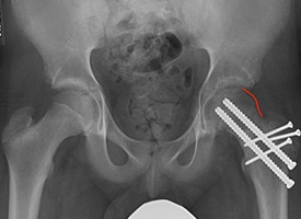
This x-ray demonstrates realignment and stabilization of the hip after surgical hip dislocation and repositioning of the severe unstable Slipped Capital Femoral Epiphysis (SCFE).
Arthroscopic Excision of a Subspine Impingement Lesion
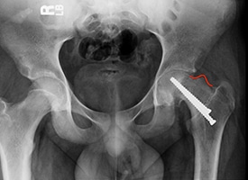
This x-ray demonstrates a subspine impingement lesion from a anterior inferior iliac spine avulsion fracture that is causing pain and decreased flexion of the hip.
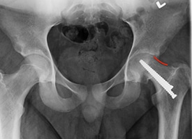
This x-ray demonstrates arthroscopic excision of a subspine impingement lesion to improve range of motion and decrease pain.
Labral Repair
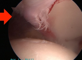
This arthroscopic photo demonstrates a labral tear.
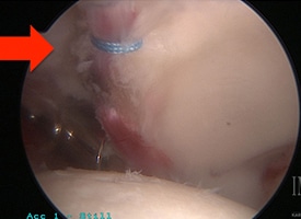
This arthroscopic photo demonstrates a labral repair.
Reshaping after Arthroscopic Re-Contouring and Labral Repair
This x-ray demonstrates femoroacetabular impingement with a CAM deformity of the femoral neck and ossification of the labrum (shown by the arrow). The deformity of the neck impinges against the socket and has causes damage to the socket.
This x-ray demonstrates reshaping of the femoral neck after arthroscopic re-contouring and labral repair. Note that the ossification of the labrum has resolved.
Reorient
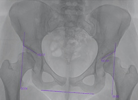
This x-ray demonstrates bilateral hip dysplasia with upward sloping hip sockets. This can cause early pain and degeneration of the hip joint.
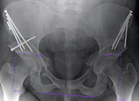
This x-ray demonstrates bilateral periacetabular osteotomies to increase coverage and stability of the hip joint. Notice that the roof of the hip socket is now horizontal, and the ball of the hip joint is now fully covered. This can decrease pain and preserve the hip joint.