Arthroscopic surgery involves placing small cameras and instruments into a joint to view, assess and treat a multitude of problems. Hip arthroscopy is a relatively new procedure unlike knee, shoulder and ankle arthroscopy. The hip is a deep, tight joint, surrounded by a thick capsule. Special instruments, positioning devices and techniques have been developed to address these conditions. With hip arthroscopy, the surgeon can view and treat many conditions that required open procedures or simply couldn’t be performed in the past. Examples include removal of loose bodies, repairing the labrum and reshaping the head and neck.

Femoral osteotomy involves cutting the thigh bone near the hip to re-align or re-direct the femoral head (ball) deeper into the acetabulum (cup). It may be performed separately or in conjunction with a pelvic osteotomy.
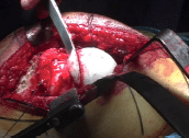
Surgical dislocation is an open dislocation of the hip. It is a relatively new procedure as well. In the past, dislocating the hip entailed a significant risk of avascular necrosis of the femoral head (AVN); damaging the blood supply to the hip. This new technique virtually eliminates that risk. Surgical dislocation can be used for all of the procedures hip arthroscopy is used for and for procedures that arthroscopy can’t be used for such as osteotomies outside of the hip joint and pathology that is too large or inaccessible with the arthroscope.
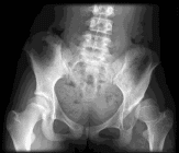
The hip is a ball and cup joint. Patients with Developmental Dysplasia of the Hip (DDH) have a shallow acetabulum (cup). The femoral head (ball) is not covered enough, which puts a lot of pressure on a small area of the acetabulum leading to pain and early arthritis. Osteotomies are used to correct these problems by directing the ball deeper into the cup, moving the cup over the ball or both.

Pelvic osteotomy involves cutting the bones in the pelvis to redirect the acetabulum (cup) over the femoral head (ball). There are many types of pelvic osteotomy. The type of osteotomy used depends on the patients’ age and the amount of correction needed.