Legg-Calve-Perthes is a temporary condition in children characterized by a transient loss of blood supply to the hip. Without an adequate blood supply, the rounded head of the femur collapses. The area can become intensely inflamed and irritated.
Perthes disease is typically seen in children between four years and ten years of age, but may be present in younger children. It is five times more common in boys than in girls. If signs and symptoms develop at a later age (>six years old) the condition can be more serious, leading to early arthritis in the hip joint.
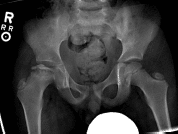
Perthes Disease occurs at the head of the femur (thigh bone). Without enough blood supply to the area, the head of the femur will collapse and become flat. Typically only one hip is affected, although it can occur on both sides.
Symptoms of Legg-Calve-Perthes disease can be intermittent over a period of weeks or even months. Pain sometimes is caused by muscle spasms that may result from irritation around the hip. Pain may be felt in other parts of the leg, such as the groin, thigh, or knee. When the hip is moved, the pain worsens. Rest often relieves the pain, as well as anti-inflammatory medication.
Each child is different, and your physician will suggest the best treatment option for your child. These include:
For children who show mild changes on their initial x-rays, treatment is usually simply observation. Observation includes regular follow-up with your pediatric orthopedist for radiographs to monitor any changes.
Taking anti-inflammatory medicine or NSAIDS (non-steroidal anti-inflammatory drugs) such as Motrin, Advil, Naproxen or Aleve as directed by your doctor may be helpful. This medication should be taken for 10 to 14 days to allow the medicine to build to therapeutic levels in the body. Taking the medication infrequently allows the medicine levels to drop, which decreases its effectiveness.
Therapy can help the patient retain or regain good range of motion of the affected hip, and can involve treatment such as hip abduction, rotation, and traction. Home traction is sometimes recommended as well.
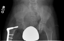
Surgery is reserved for the most severe cases and is recommended to decrease long term damage to the hip. Surgical treatment re-establishes the proper align-ment of the bones of the hip. The head of the femur is placed deep within the socket, or acetabulum. This alignment is kept in place with screws and plates, which will be removed at a later time. In some cases, the socket must also be made deeper because the head of the femur has actually enlarged during the healing process and no longer fits snugly within. After either procedure, the child is often placed in a cast from the chest to the toes for 6 to 8 weeks.
After the cast is removed, the child will again participate in physical therapy. Activities will be designed so that the child only partially bears weight on the affected hip until it has healed. X-rays will show when the final stages of the healing are under way.