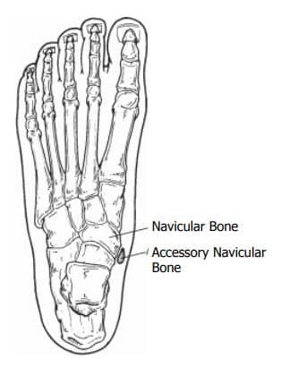
An accessory navicular bone is a congenital abnormality, meaning it was present since birth. It is not part of normal bone structure and therefore not found in most people.
An extra bone develops in the posterior tendon in the foot (see diagram below) at the insertion (this is where the bone attaches to the muscle) onto the tarsal navicular bone. The accessory navicular bone occurs alongside the navicular bone and is typically attached to normal bone through a cartilage structure similar to a growth plate (the ends of long bones in children).
Treatment for an accessory navicular can include anti-inflammatory medication, resting the affected foot, applying a cat, or immobilization in a CAM boot. Stretching and physical therapy may also help. Surgery is recommended when other treatment options fail. Each child is different, and your physician will suggest the best treatment option for your child.
Taking anti-inflammatory medicine or NSAIDS (non-steroidal anti-inflammatory drugs) such as Motrin, Advil, Naproxen or Aleve as directed by your doctor can be effective. This medication should be taken for 10 to 14 days to allow the medicine to reach therapeutic levels in the body.
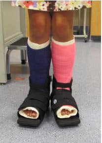
A short-leg cast (a cast that is applied below the knee) is usually worn for 3 to 6 weeks. After the cast is removed, some patients are prescribed arch supports or custom insoles which fit into their shoes. Stretching exercises are prescribed after cast removal.
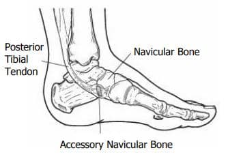
If all other treatment fails to relieve the symptoms of accessory navicular syndrome, surgery may be necessary. This bone is not needed for normal foot function, so there is no negative effect if it is removed. Surgery involves removing the accessory navicular bone, reshaping the edge of the remaining navicular, and repairing the posterior tibial tendon to improve its function.