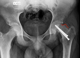
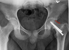
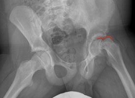
Specialists in Pediatric Hips
The Hip Disorders Program at Children’s Orthopaedic’s specializes in diagnosis, treatment and long-term care for infants, children, teens and young adults with congenital, developmental and post-traumatic hip disorders.
Whether your child is a premature infant with a congenital hip dislocation, an adolescent with Legg-Calve-Perthes diseases, a teen with hip pain during sports, or a young adult with hip dysplasia, our specialty-trained pediatric orthopedic team has the expertise needed to produce the best possible outcomes.
Our team considers all possible treatment options for children, adolescents and young adults with hip disorders. We use a full spectrum of diagnostic testing and have access to the expertise and full resources of Johns Hopkins All Children’s Hospital in every pediatric subspecialty. Along with standard imaging technologies such as x-rays, CT scans, and MRI’s, the Hip Disorders Program at Children’s Orthopaedics also uses disease-specific MRI protocols and 3D CT reconstructions to help guide treatment. With better imaging of the hip, our physicians can provide a more accurate diagnosis and help guide surgical treatment if needed.
Empowering Active Futures
What You Need to Know
Many teens and young adults suffer from variety of hip disorders and conditions that cause structural abnormalities of the hip joint. If left untreated, these often-painful conditions can lead to early degeneration and premature osteoarthritis of the hip joint in otherwise young, healthy, and active individuals. Once severe degeneration and osteoarthritis has occurred, hip replacement is often the only solution.
Hip replacement is surgically removing the natural hip joint you were born with and replacing it with a metal and plastic hip joint. Although a hip replacement has an excellent outcome, there is concern for the longevity of the artificial joint in the young and active patient. A young patient with a hip replacement will often need multiple revision joint replacement surgeries throughout their life. Therefore, it is necessary to avoid or postpone hip joint replacement.
Hip preservation focuses on treatment strategies to correct any structural abnormalities of the hip in order to decrease pain and halt progression of hip degeneration or osteoarthritis in young patients with congenital, developmental, and post-traumatic hip disorders.
As our understanding of structural abnormalities in the hip has increased, our specialists have been able to better identify underlying conditions in earlier stages, providing patients with more treatment options that help to preserve the integrity of the hip joint, including both arthroscopic and open surgical techniques.
Re-contouring or reshaping complex abnormalities of the hip joint can restore natural range of motion and prevent arthritis.
PRE OP
The red line demonstrates a large residual CAM deformity after in situ screw fixation of Slipped Capital Femoral Epiphysis (SCFE). This deformity limits range of motion and impinges against the hip socket causing damage to the joint.
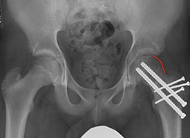
POST OP
The red line demonstrates re-contouring of the femoral neck after arthroscopic treatment. This treatment increases range of motion and prevents further damage to the hip.
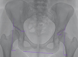
Reorienting or repositioning a shallow hip socket can restore the natural physiologic position to decrease shear forces in the hip joint and prevent arthritis.
PRE OP
This x-ray demonstrates bilateral hip dysplasia with upward sloping hip sockets. This can cause early pain and degeneration of the hip joint.
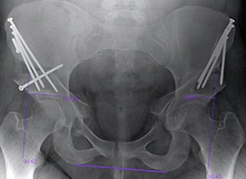
POST OP
This x-ray demonstrates bilateral periacetabular osteotomies to increase coverage and stability of the hip joint. Notice that the roof of the hip socket is now horizontal, and the ball of the hip joint is now fully covered. This can decrease pain and preserve the hip joint.
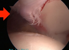
Repairing the labrum, cartilage, and other soft tissues around the hip joint can restore function and preserve longevity of the joint.
PRE OP
This arthroscopic photo demonstrates a labral tear.
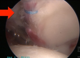
POST OP
This arthroscopic photo demonstrates a labral repair.
Realignment of some hip conditions can restore the normal anatomy to preserve the hip joint.
PRE OP
This x-ray demonstrates a severe unstable Slipped Capital Femoral Epiphysis (SCFE) of the left hip.
POST OP
This x-ray demonstrates realignment and stabilization of the hip after surgical hip dislocation and repositioning of the severe unstable Slipped Capital Femoral Epiphysis (SCFE).
Board Certified Orthopaedic Surgeon
Specialized Care for Young Hips
Dr. Drew Warnick and Dr. Daniel Bland are trained in specialized hip preservation techniques that are only available at a few centers on the entire west coast of Florida.
These techniques include surgical dislocation of the hip, the Ganz Periacetabular Osteotomy, and surgical head realignment after Slipped Capital Femoral Epiphysis. Our physicians are also trained in minimally invasive surgical techniques such as hip arthroscopy, which can sometimes reduce the need for more extensive and invasive surgeries.